Aim:
Corneal neovascularization is a process of pathological ingrowth of blood vessels from the limbal vascular plexus into the cornea, which occurs e.g. due to corneal hypoxia, corneal infection, trauma, and other immunological processes. This work aimed to develop an application using machine learning to detect corneal blood vessels and determine their quantity automatically.
Methodology:
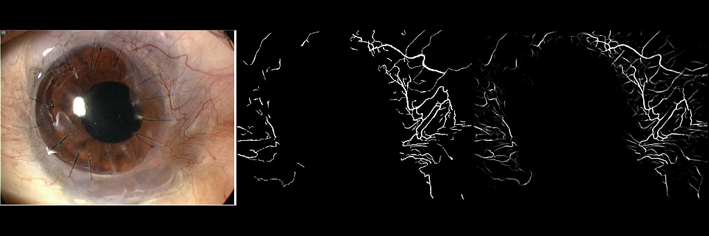
Two phases were used to create the software application. In the first computational phase, a neural network was trained. The network can identify the corneal vessels in the eye image, which is processed, and then quantification is performed. In the second phase, the user interface was created, which serves the user (doctor) and where the individual images can be viewed and compared in detail.
Results:
The result of the project is a software application that can automatically assess the presence of pathological corneal neovascularisation and quantify it based on the provided images. The user (doctor) can view, compare over time and evaluate the images in a simple user interface.
Conclusion:
Corneal neovascularizations accompanying many corneal diseases may occur asymptomatically in the patient but may contribute to a large extent to the reduction of corneal transparency and, thus, impairment of visual function. Until now, the presence and development of corneal neovascularization could only be monitored manually with the naked eye of the physician using slit-lamp images. According to the authors, an automated application that recognizes corneal vessels and enables their quantification could help physicians more accurately monitor this pathology’s status and development and better target and monitor treatment.